
TL;DR

Prime made for students and young adults
- Fast, free delivery for dorm and study essentials
- Prime Video and Amazon Music included
- Member-only deals
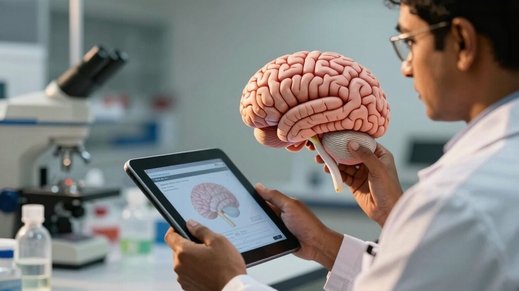
Indian scientists have developed the most detailed 3D atlas of the human brainstem to date. This breakthrough enhances understanding of brain anatomy and could impact neurological research and treatment.
Indian scientists have unveiled the most detailed three-dimensional (3D) atlas of the human brainstem to date, representing a notable development in neuroscience research. This development, announced in March 2024, provides detailed insights into the complex structure of this brain region, which is involved in regulating functions such as breathing, heart rate, and consciousness.
The team, led by researchers at the Indian Institute of Neurosciences and Brain Research, used advanced imaging techniques and computational modeling to construct a detailed 3D map of the human brainstem. The atlas incorporates thousands of high-resolution images, enabling visualization of neural pathways, nuclei, and fiber tracts within the brainstem.
According to the lead researcher, Dr. Anjali Kumar, this atlas offers improvements in resolution and detail over previous models. “Our work provides an anatomical reference that can support clinical diagnosis and neuroscientific research,” she stated. The project involved collaboration across multiple disciplines, including neuroimaging, bioinformatics, and neuroanatomy.
While the atlas is still undergoing validation, initial feedback from the scientific community has highlighted its potential to aid in understanding brainstem-related disorders such as stroke, Parkinson’s disease, and multiple sclerosis. The team plans to make the atlas publicly accessible for researchers worldwide in the coming months.
Implications for Neuroscience and Medical Research
This development is relevant because the brainstem is a vital and complex part of the brain, involved in regulating essential functions. A detailed 3D atlas can assist researchers and clinicians in understanding its anatomy, which may support the development of diagnostic tools and targeted treatments for neurological conditions. The atlas could also serve as a resource for future studies on brain connectivity and function.
Additionally, this achievement positions India as a contributor to neuroimaging and brain mapping efforts, providing data that may be useful in the global neuroscience community. It may also support the development of surgical techniques and neuroprosthetics that require precise anatomical information.
As an affiliate, we earn on qualifying purchases.
Advances in Brain Mapping and Indian Neuroscience Efforts
Recent years have seen increased efforts worldwide to improve brain mapping technologies, driven by advances in imaging and computational analysis. Historically, detailed brain atlases have primarily focused on the cerebral cortex, with fewer comprehensive models of the brainstem due to its size and structural complexity.
India has invested significantly in neuroscience research, with institutions such as the Indian Institute of Neurosciences and Brain Research leading innovative projects. This new atlas builds on previous efforts but distinguishes itself through its high resolution and detailed representation of neural pathways within the brainstem, a region essential for survival and less extensively mapped.
Earlier models of the brainstem were often based on post-mortem dissections or lower-resolution imaging, limiting their ability to capture the full complexity of neural connections. The new 3D atlas aims to address these limitations by providing a more comprehensive and accurate anatomical reference.
“This atlas offers a detailed anatomical reference that can support clinical diagnosis and neuroscientific research.”
— Dr. Anjali Kumar, lead researcher
As an affiliate, we earn on qualifying purchases.
Validation and Practical Applications Still Under Development
Although the atlas has been announced and initial feedback has been positive, it is still undergoing validation processes to confirm its accuracy and utility. The extent to which it will be adopted in clinical settings or influence surgical planning remains to be determined. Further research and validation studies are necessary to assess its potential impact on understanding brainstem diseases.
It is also uncertain how quickly the atlas will be integrated into medical practice or how it compares to existing models in practical applications.
neuroscience research anatomical models
As an affiliate, we earn on qualifying purchases.
As an affiliate, we earn on qualifying purchases.
Upcoming Validation, Access, and Research Opportunities
The research team plans to publish detailed validation results in peer-reviewed journals within the next few months. They also intend to release the atlas publicly for use by neuroscientists and clinicians worldwide, supporting further research and development.
Future efforts may include applying the atlas in clinical case studies, refining it with additional data, and exploring its use in surgical planning and neuroprosthetics. The team also plans to collaborate with international institutions to expand the atlas’s applications.
As an affiliate, we earn on qualifying purchases.
Key Questions
How does this new atlas differ from previous brainstem maps?
The new atlas offers higher resolution and detailed visualization of neural pathways, nuclei, and fiber tracts compared to earlier models based on dissection or lower-resolution imaging.
When will the atlas be available for public use?
The research team plans to release the atlas publicly within the next few months after completing validation and peer review processes.
What potential medical benefits does this development offer?
It could improve diagnosis and treatment of brainstem-related disorders, support surgical planning, and facilitate neuroresearch by providing a detailed anatomical reference.
Is this development unique to India?
While many countries are involved in brain mapping research, this high-resolution 3D brainstem atlas is a notable achievement by Indian scientists, contributing to global efforts in neuroanatomy.
Source: hn
Fall Picks
fall essentials
As an affiliate, we earn on qualifying purchases.